Region
Hip
The deep socket that carries your weight to your legs, and the muscles that extend, rotate and steady it.
10 structures · 8 muscles · 2 joints
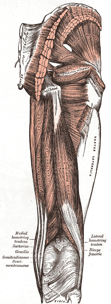
The gluteal region: gluteus maximus and medius over the pelvis, with the hamstrings running down behind the thigh.
The plate is captioned in the book, but that caption did not come through the scan legibly, so this description was written here from looking at the plate rather than guessed at.
Henry Vandyke Carter, plate 434 of Anatomy of the Human Body (Henry Gray, 1918). Public domain — file on Wikimedia Commons.

Bone structure
- Ilium
- Ischium
- Pubis
- Sacrum
- Femur (upper end)
Key functions
- Hip extension
- Hip external rotation
- Hip abduction
- Pelvic stabilisation
- Hip internal rotation
- Hip flexion
- Hip adduction
- Hip rotation
- Circumduction
- Lumbar stabilisation
- Load transfer
Muscle structure
- Gluteus maximus hip extension · hip external rotation
- Gluteus medius hip abduction · pelvic stabilisation · hip internal rotation · hip external rotation
- Gluteus minimus hip abduction · pelvic stabilisation · hip internal rotation
- Iliacus hip flexion
- Iliopsoas hip flexion · lumbar stabilisation
- Piriformis hip external rotation · hip abduction
- Psoas major hip flexion · lumbar stabilisation
- Tensor fasciae latae hip flexion · hip abduction · hip internal rotation
Joints
- Hip hip flexion · hip extension · hip abduction · hip adduction · hip rotation · circumduction
- Sacroiliac load transfer
Tendons and attachments
- Gluteus maximus
- from Posterior gluteal line of the ilium, the back of the sacrum and coccyx, the sacrotuberous ligament and the thoracolumbar fascia
- to Mostly the iliotibial tract; the deeper lower fibres onto the gluteal tuberosity of the femur
- Gluteus medius
- from Outer surface of the ilium, between the anterior and posterior gluteal lines
- to Lateral surface of the greater trochanter of the femur
- Gluteus minimus
- from Outer surface of the ilium, between the anterior and inferior gluteal lines
- to Anterior border of the greater trochanter of the femur
- Iliacus
- from Upper two-thirds of the iliac fossa — the broad inner hollow of the pelvic bone
- to Lesser trochanter of the femur, via the common iliopsoas tendon
- Iliopsoas
- from Two separate origins — the lumbar spine (psoas major) and the inner surface of the ilium (iliacus)
- to Lesser trochanter of the femur, by a common tendon
- Piriformis
- from Anterior surface of the sacrum, around the second to fourth sacral segments
- to Upper border of the greater trochanter of the femur
- Psoas major
- from Bodies and transverse processes of the twelfth thoracic and all lumbar vertebrae, and the discs between them
- to Lesser trochanter of the femur, via the common iliopsoas tendon
- Tensor fasciae latae
- from Anterior superior iliac spine and the adjacent outer lip of the iliac crest
- to The iliotibial tract, about a third of the way down the thigh
Movements that load it
Where this came from
Every name, attachment and action on this page is read from The Atlas’s own records — each one checked against Terminologia Anatomica, and each one linked above so you can read the full entry. Nothing here is retyped or approximated, so this page cannot drift from the collection: it is the collection, arranged by region.
The figure is different in kind: it is sourced, not drawn here and not generated. A diagram is a claim, and an invented one is a claim nobody can check — so the plates are Henry Vandyke Carter’s, engraved for the 1918 edition and long out of copyright, credited above with a link to the file so you can verify the licence yourself. Being 1918, they carry the anatomical names of their day, which do not always match the modern ones used in the records.
Plate captions were read from the Internet Archive’s scan of the 1918 edition (anatomyofhumanbo00grayrich), and the licence of every plate was checked against its own file record on Commons rather than assumed from the age of the book. The build refuses to publish a page whose plate is not public domain.