Region
Thigh
The largest muscle mass in the body. Everything that straightens your knee, and most of what bends it.
13 structures · 13 muscles
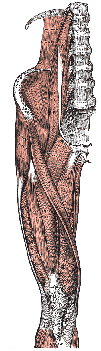
Muscles of the iliac and anterior femoral regions.
That is the plate’s own caption, as printed in the 1918 edition.
Henry Vandyke Carter, plate 430 of Anatomy of the Human Body (Henry Gray, 1918). Public domain — file on Wikimedia Commons.

Bone structure
- Femur
- Patella
Key functions
- Hip adduction
- Hip flexion
- Hip extension
- Knee flexion
- Knee external rotation
- Knee internal rotation
- Knee extension
- Hip abduction
- Hip external rotation
Muscle structure
- Adductor longus hip adduction · hip flexion
- Adductor magnus hip adduction · hip extension
- Biceps femoris knee flexion · hip extension · knee external rotation
- Gracilis hip adduction · knee flexion · knee internal rotation
- Pectineus hip flexion · hip adduction
- Quadriceps femoris knee extension · hip flexion
- Rectus femoris knee extension · hip flexion
- Sartorius hip flexion · hip abduction · hip external rotation · knee flexion
- Semimembranosus knee flexion · hip extension · knee internal rotation
- Semitendinosus knee flexion · hip extension · knee internal rotation
- Vastus intermedius knee extension
- Vastus lateralis knee extension
- Vastus medialis knee extension
Tendons and attachments
- Adductor longus
- from Front of the body of the pubis, just below the pubic crest
- to Middle third of the linea aspera of the femur
- Adductor magnus
- from Inferior pubic ramus, ramus of the ischium, and the ischial tuberosity
- to The linea aspera and medial supracondylar line of the femur, and the adductor tubercle
- Biceps femoris
- from Long head from the ischial tuberosity; short head from the lateral lip of the linea aspera of the femur
- to Head of the fibula, with a slip to the lateral condyle of the tibia
- Gracilis
- from Lower half of the body of the pubis and the inferior pubic ramus
- to Upper medial surface of the tibia, as part of the pes anserinus
- Pectineus
- from Pecten pubis — the ridge on the superior ramus of the pubis
- to Pectineal line of the femur, between the lesser trochanter and the linea aspera
- Quadriceps femoris
- from Four separate heads — one from the pelvis, three from the shaft of the femur
- to The tibial tuberosity, via the quadriceps tendon, the patella and the patellar ligament
- Rectus femoris
- from Two heads — the anterior inferior iliac spine (straight head) and the groove above the acetabulum (reflected head)
- to Base of the patella, then the tibial tuberosity via the patellar ligament
- Sartorius
- from Anterior superior iliac spine
- to Upper medial surface of the tibia, as part of the pes anserinus
- Semimembranosus
- from Ischial tuberosity, by a broad flat tendon deep to the other hamstrings
- to Posterior aspect of the medial condyle of the tibia
- Semitendinosus
- from Ischial tuberosity, by a tendon shared with the long head of biceps femoris
- to Upper medial surface of the tibia, as part of the pes anserinus
- Vastus intermedius
- from Anterior and lateral surfaces of the upper two-thirds of the shaft of the femur
- to Deep surface of the quadriceps tendon, then the patella and the tibial tuberosity
- Vastus lateralis
- from Greater trochanter, the intertrochanteric line and the lateral lip of the linea aspera of the femur
- to Lateral border of the patella, then the tibial tuberosity via the patellar ligament
- Vastus medialis
- from Intertrochanteric line and the medial lip of the linea aspera of the femur
- to Medial border of the patella, then the tibial tuberosity via the patellar ligament
Movements that load it
Where this came from
Every name, attachment and action on this page is read from The Atlas’s own records — each one checked against Terminologia Anatomica, and each one linked above so you can read the full entry. Nothing here is retyped or approximated, so this page cannot drift from the collection: it is the collection, arranged by region.
The figure is different in kind: it is sourced, not drawn here and not generated. A diagram is a claim, and an invented one is a claim nobody can check — so the plates are Henry Vandyke Carter’s, engraved for the 1918 edition and long out of copyright, credited above with a link to the file so you can verify the licence yourself. Being 1918, they carry the anatomical names of their day, which do not always match the modern ones used in the records.
Plate captions were read from the Internet Archive’s scan of the 1918 edition (anatomyofhumanbo00grayrich), and the licence of every plate was checked against its own file record on Commons rather than assumed from the age of the book. The build refuses to publish a page whose plate is not public domain.