Region
Leg
Below the knee: the calf that pushes you off the ground, and the muscles that steer and steady your foot.
11 structures · 9 muscles · 2 joints
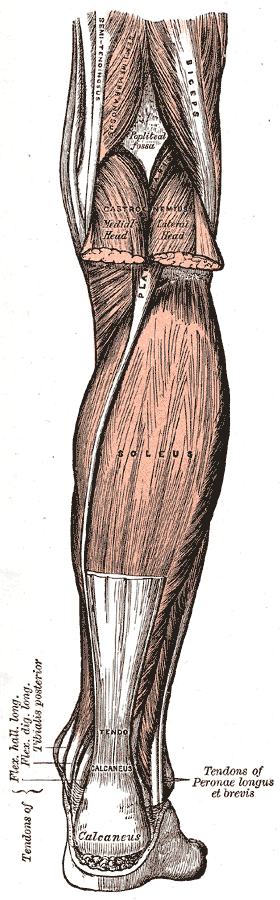
Muscles of the back of the leg.
That is the plate’s own caption, as printed in the 1918 edition.
Henry Vandyke Carter, plate 438 of Anatomy of the Human Body (Henry Gray, 1918). Public domain — file on Wikimedia Commons.

This plate labels the tendons behind the ankle Peronæi longus et brevis. The records here call those muscles fibularis longus and fibularis brevis, which is the name Terminologia Anatomica uses. Same two muscles, a century apart.
Bone structure
- Tibia
- Fibula
Key functions
- Ankle dorsiflexion
- Ankle plantarflexion
- Foot eversion
- Knee flexion
- Knee extension
- Knee rotation
- Knee internal rotation
- Foot inversion
Muscle structure
- Fibularis brevis foot eversion · ankle plantarflexion
- Fibularis longus foot eversion · ankle plantarflexion
- Gastrocnemius ankle plantarflexion · knee flexion
- Plantaris ankle plantarflexion · knee flexion
- Popliteus knee flexion · knee internal rotation
- Soleus ankle plantarflexion
- Tibialis anterior ankle dorsiflexion · foot inversion
- Tibialis posterior foot inversion · ankle plantarflexion
- Triceps surae ankle plantarflexion · knee flexion
Joints
Tendons and attachments
- Fibularis brevis
- from Lower two-thirds of the lateral surface of the fibula
- to Tuberosity at the base of the fifth metatarsal
- Fibularis longus
- from Head and upper lateral surface of the fibula
- to Medial cuneiform and the base of the first metatarsal, crossing the sole of the foot
- Gastrocnemius
- from Medial and lateral condyles of the femur, above the knee
- to Calcaneal tuberosity, via the calcaneal tendon
- Plantaris
- from Lateral supracondylar line of the femur, above the lateral head of gastrocnemius
- to Calcaneal tuberosity, medial to the calcaneal tendon
- Popliteus
- from Lateral condyle of the femur, within the knee joint capsule
- to Posterior surface of the tibia, above the soleal line
- Soleus
- from Posterior surfaces of the head and upper shaft of the fibula, and the soleal line of the tibia
- to Calcaneal tuberosity, via the calcaneal tendon
- Tibialis anterior
- from Lateral condyle and upper lateral surface of the tibia, and the interosseous membrane
- to Medial cuneiform and the base of the first metatarsal
- Tibialis posterior
- from Interosseous membrane and the adjacent posterior surfaces of the tibia and fibula
- to Navicular tuberosity, with slips to the cuneiforms, cuboid and middle metatarsals
- Triceps surae
- from Three heads — two from the femoral condyles (gastrocnemius), one from the tibia and fibula (soleus)
- to Calcaneal tuberosity, via the calcaneal tendon
Movements that load it
Where this came from
Every name, attachment and action on this page is read from The Atlas’s own records — each one checked against Terminologia Anatomica, and each one linked above so you can read the full entry. Nothing here is retyped or approximated, so this page cannot drift from the collection: it is the collection, arranged by region.
The figure is different in kind: it is sourced, not drawn here and not generated. A diagram is a claim, and an invented one is a claim nobody can check — so the plates are Henry Vandyke Carter’s, engraved for the 1918 edition and long out of copyright, credited above with a link to the file so you can verify the licence yourself. Being 1918, they carry the anatomical names of their day, which do not always match the modern ones used in the records.
Plate captions were read from the Internet Archive’s scan of the 1918 edition (anatomyofhumanbo00grayrich), and the licence of every plate was checked against its own file record on Commons rather than assumed from the age of the book. The build refuses to publish a page whose plate is not public domain.